Stool microscopic examination images pdf
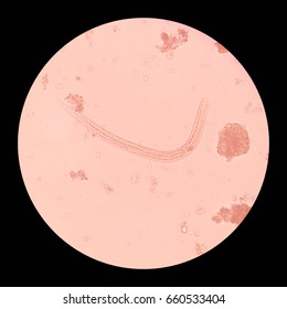
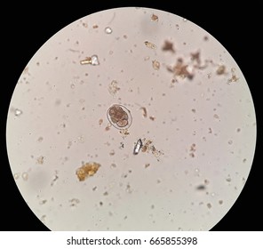
1/10/2018 · Stool examination (see the images below) for trophozoites or cysts is the traditional method for diagnosing giardiasis. At least 3 stools taken at 2-day intervals should be examined for ova and parasites. Trophozoites may be found in fresh, watery stools but disintegrate rapidly. If the stool is not fresh or is semiformed to formed, trophozoites will not be found.
Using the Next button (below) you’ll find a total of 13,580 Stool images for you to choose from! Or use the search tool above to find other images illustrating almost anything you can imagine. And at any time you can click on any thumbnail pic you see to enlarge it. Then, if you like what you see, click again to buy it then instantly download it.
Routine Stool Analysis Sr. No. Observation Normal Reading Conclusion A Macroscopic Examination Normal Color Consistency Solid, semi solid
Microscopic colitis is an inflammation of the colon that a health care provider can see only with a microscope. Inflammation is the body’s normal response to injury, irritation, or infection of tissues. Microscopic colitis is a type of inflammatory bowel disease—the general name for diseases
Most intestinal infections resolve within a few days of onset, and microscopic evaluation of stool for parasites in such short-term enteritis is usually unrevealing. The approach to organism identification via stool microscopy should be guided by the clinical presentation, including geographic exposure, age, and nature of clinical symptoms.
Stool microscopy is a diagnostic tool for identification of parasitic organisms including protozoa and helminths; it is also useful for quantification of fecal leukocytes. Protozoa are represent one group within the kingdom Protista; other Protista include protophyta and certain molds.
Stool Routine Examination howMed

Parasites Complete Microscopic Examination Stool
Examination findings are unremarkable: dehydration is rare and the rectal mucosa looks normal on sigmoidoscopy. Stool analysis should be undertaken to rule out infection but is negative for pathogens in microscopic colitis. Blood test abnormalities are rare, but coeliac serology should be checked. Colonoscopic macroscopic abnormalities have been suggested in up to 30% of cases but are subtle
This chapter discusses about macroscopic examination and microscopic examination of fecal specimens. The microscopic examination of the stool specimen, normally called the ova and parasite examination, consists of three separate techniques: the direct wet smear, the concentration (sedimentation and flotation), and the permanent stained smear.
Stool analysis allows examination of faeces (stools). Stool analysis is useful for solving stomach, bowel or other digestive problems. Stool analysis is useful for solving stomach, bowel or …
Importance of microscopic stool examination in patients with diarrhoea. Shah SA, Marwat SA, Rashid HU, Hussain A, Khurshid K, Ahmad S. BACKGROUND: Loose motion is a common symptom in patients reporting to our hospital.
Your doctor can order a stool ova and parasites (O&P) test to check for parasites and their eggs in your stool, or feces. It’s a relatively easy and common test.
The mucus present in the stool is the one that should be protecting the intestinal lining. The lining is left exposed after the mucus is eaten away. It is therefore noted that presence of mucus in your stool warrants that more problems are yet to come. Once the mucosal layer is worn out, pathogens can then easily find their way into the body. If left untreated, this may lead to even more
PROCEDURE MANUAL MOUNT SINAI HOSPITAL/TORONTO MEDICAL LABORATORIES SHARED MICROBIOLOGY SERVICE Page 1 MSH/TML Shared Microbiology Service Policy & Procedure Manual
Microscopy is the technical field of using microscopes to view objects and areas of objects that cannot be seen with the naked eye (objects that are not within the resolution range of the normal eye). There are three well-known branches of microscopy: optical, electron, and scanning probe microscopy, along with the emerging field of X-ray
microscopic chart or reference book pictures. White blood cells, red blood cells, epithelial cells, White blood cells, red blood cells, epithelial cells, bacteria, various types of casts and crystals that may be encountered should be included as well as
Purpose and Scope: The microscopic examination may be used to determine the presence of leukocytes and erythrocytes in a fecal smear. This will very quickly give the clinician information on the patients disease state.
27/04/2014 · In this video we discuss how to perform the microscopic examinations of stool sample in parasitology lab.
Parasafe Pinworm Collectors paddle viewed directly on microscope stage The clearer and thinner plastic paddle on this Pinworm Collector makes it
STOOL EXAMINATION 1. Guided by Presented by Dr. R K Hibare Dr. Madhusudan B G Prof & HOD, Dept of Roga Nidana I MD, Dept of Roga Nidana GAMC, Bengaluru GAMC, Bengaluru
investigating the presence of yeast, disparity may exist between culturing and microscopic examination. Yeast are not uniformly dispersed throughout the stool and this may lead to undetectable or low levels of yeast identified by microscopy, despite culture and identified yeast species. Conversely, microscopic examination may reveal a significant amount of yeast present but no viable yeast

The classical technique described for the microscopic examination of parasites is the iodine mount method, which aids in the differentiation and identification of parasites by characteristic morphological features and details of internal structures. The method is simple to perform, quick, and inexpensive, facilitating direct visualization of parasitic ova and cyst morphology. The disadvantage
Laboratory Guidelines For Intestinal Parasites Suggested Physician Ordering Plan for the Laboratory Examination Of Stool Specimens Washington State Clinical
Adjustable Height Bar Stool (Set of 2) by Creative Images International If you want to buy Adjustable Height Bar Stool (Set of 2) by Creative Images International Ok you want deals and save. online looking has now gone an extended method; it has changed the …
Microscopic Examination Microstructure Analysis to Evaluate Materials. During Microstructure Analysis of metals and alloys, a Microscopic Examination is conducted to study the structure of the material under magnification.
Lab Dept: Microbiology/Virology Test Name: OVA AND PARASITE EXAMINATION, STOOL General Information Lab Order Codes: Test Includes: Examination of stool for intestinal parasites by direct/concentrated microscopic exam and trichrome stain. This test does not include: Cryptosporidium, Cyclospora, Microsporidium or Pinworm. If only Cryptosporidium or Giardia are requested, refer to …
These images are provided for use as teaching aids for the training and education of clinical laboratory scientists. The images cannot be downloaded and used without my consent.
Intussusception is a medical condition in which a part of the intestine folds into the section next to it. It typically involves the small bowel and less commonly the large bowel. Symptoms include abdominal pain which may come and go, vomiting, abdominal bloating, and bloody stool. It often results in a small bowel obstruction. Other
Routine Ova and Parasite Exam State Public Health
Exam the stool before giving antibiotic or other drugs. Semi-formed stool should be examined within 60 minutes of collection. Liquid stool should be examined within the first 30 minutes. Solid stool should be examined within the first hour of collection. Trophozoites degenerate in liquid stool rapidly, so exam the stool within 30 minutes. In the case of constipated cases, use non-residual
A freshly passed stool is the specimen of choice. Stool specimen should be collected before antibiotic therapy, or as early in the course of the disease. If blood or mucous is present, it should be included in the specimen Patients receiving tetracyclines, anti-diarrheal drugs, barium, bismuth, oil, iron , or magnesium may not yield accurate results. 2. Bismuth found in toilet tissue
Appearance of blood in stools. How the blood looks depends on where it is coming from. Spots of red blood on the toilet paper, drops in the toilet bowl or blood on the surface of your stool indicate a problem in the anus and lower rectum.
microscopic examination. Yeast are not uniformly dispersed throughout the stool, this may lead to Yeast are not uniformly dispersed throughout the stool, this may lead to undetectable or low levels of yeast identified by microscopy, despite a cultured amount of yeast.
Download PDF Info Publication number US5874315A. US5874315A mercury based fixatives have been used to preserve stool specimens for staining and microscopic parasitological examination. Stool parasites that have been preserved in mercury based fixatives provide good definition of the parasite upon staining. However, mercury based fixatives have a number of disadvantages … – python extract images from pdf Find stool Stock Images in HD and millions of other royalty-free stock photos, illustrations, and vectors in the Shutterstock collection. Thousands of new, high-quality pictures added every day.
Normally, stool is colored in light to dark brown color. The normal color of the stool comes from chemical substance-bilirubin, which is produced by liver from hemoglobin, after red blood cells have finished their life cycle and die.
These images are a random sampling from a Bing search on the term “Parasite Examination of the Stool.” Click on the image (or right click) to open the source website in a new browser window.
Objective: To compare the routine and concentration techniques on microscopic examination of stool for parasitic ova and cysts to emphasize importance of concentration technique Methods: A descriptive study of 100 symptomatic and asymptomatic patients who attended the Microbiology
know how much microscopic stool examination can help us in management of patients with diarrhoea. Methods: This cross-sectional descriptive study was conducted from January 2010 to April 2012, at Thall Scout Hospital, Hangoo, Khyber Pukhtoon Khwa, Pakistan.
ersion – eectie date 00206 microscopy examination of thick and thin blood films for identification of malaria parasites malaria microscopy standard operating procedure – mm-sop-08
Objective : To perform Microscopic examination of Stool Requirement : Glass slides, Cover slips, Normal saline, lugol’s Iodine Solution, Specimen : Morning stool specimen collected in a wide mouth clean & dry container Pre-Analytical Variables : Specimen collection , Labelling of specimen, Name, Identifying number, date, time Procedure : Saline specimen Preparation Place a drop of normal
Microscopic examination of stool specimens is the cornerstone of detection of intestinal parasites in parasitology laboratories. In Europe, fresh, nonpreserved stool specimens are generally used
microscopic examination of chancre fluid, lymph node aspirates, blood, bone marrow 50,000 to 70,000 people; only found in Africa tsetse fly , day-biting fly of the genus Glossina
If a complete microscopic exam is desired on this specimen, call 314-362-3898 to request complete microscopic exam. Collection Procedure: Using a Parasitology Transport Kit, collect stool as described in instructions provided with each kit.
The routine Ova and Parasite exam for intestinal parasites includes a microscopic examination of a concentrated wet mount (using the stool collected in the 10% formalin vial), a trichrome stained slide (using the stool collected in the PVA vial), and the Meridian Merifluor DFA for Cryptosporidium and Giardia (using the stool collected in the 10% formalin vial).
Download stunning free images about Stool. Free for commercial use No attribution required
zdescribe microscopic examination of stool zexplain the chemical examination of stool zdescribe the preservation of stool 50.2 PHYSICAL EXAMINATION OF STOOL The following aspects of stool should be examined (a) Quantity: In intestinal amoebiasis the stools tend to be voluminous. Whereas in bacillary dysentery due to Shigella the stools are scanty in quantity (b) Consistency and form: Normal
Macroscopic examination of stool sample: The stool sample is checked for its labeling and identified. Macroscopic features such as consistency, evidence for blood and mucus, larval or adult forms is noted.
microscopic images are previously segmented to separate the parasite image from the background. The extracted parasite is then resized to 12×12 image features vector. For dimensionality reduction, the principal component analysis basis projection has been used. 12×12 extracted features were orthogonalized into two principal components variables that consist the input vector of the PNN. The …
for detection of Giardia lamblia antigen in stool. Of 84 stool samples from children in Duhok governorate, 42 Of 84 stool samples from children in Duhok governorate, 42 were positive and 42 negative for G. lamblia or other parasites by direct and indirect microscopic examination.
The microscopic examination of the centrifuged urine sediment includes the study of formed elements, such as WBC’s, RBC’s, casts and crystals. The macroscopic examination of urine includes physical appearance, such as color, character and clarity.
Transport Time relapse before processing the sample Stool samples should be examined and cultured as soon as possible after collection. As the stool
Stool examination Stool analysis determines the various properties of the stool for diagnostic purposes. Frequently ordered tests on faeces includes tests for leukocytes, blood, fat, parasites, and pathogens. Bacteria, viruses, intestinal parasites and other malfunctions can be revealed from stool samples. Stool cultures are necessary in epidemiology and public health studies. Microscopic exam
Stool Analysis What is the used 200-250 mOsm with serum PPT. Presentation Summary : Stool Analysis What is the used 200-250 mOsm with serum osmol- arity to calculate osmotic gap Sodium 5.8-9.8 mEq / 24hr Chemical examination Normal values
Manual Microscopic Urinalysis Exam pSMILE Portal
To maximize recovery of cysts, stool samples in formalin, or other fixatives, should be con centrated prior to microscopic examination (e.g., 10 min at 500 × g when using the forma lin-ethyl acetate concentration procedure).
Microscopic Examination of Feces Print; Details Hits: 40362 Page 1 of 2. Cells. Pus Cells Normally, a few are present. More than a few indicates bacillary dysentery or ulcerative colitis. Epithelial Cells Normally a few are present. If many are present, however, it indicates inflammation of the bowels. Macrophages Normally, present only occasionally. If many are present, it indicates bacillary
If stool is black in color, there is a chance of upper GI bleeding. Frank blood (fresh) in the stool gives an indication of lower GI bleeding and hemorrhoids. Frank blood (fresh) in the stool gives an indication of lower GI bleeding and hemorrhoids.
Methods: Microscopic examination (Auramin stain confirmed by Kinyoun stain), Crypto-strip (Coris Bioconcept), ELISA (Novitec Cryptosporidium ELISA) and …
Abstract. Microscopic examination of stool samples from captive nonhuman primates with diarrhea for the presence or absence of leukocytes or erythrocytes or both as a means of predicting the presence of Shigella spp. was performed.
Microscopic examination of stools from nonhuman primates

Stool Analysis microscopy.ahs.chula.ac.th
Download stool test stock photos. Affordable and search from millions of royalty free images, photos and vectors.
Detection of Giardia lamblia antigen in stool specimens

Microscopic examinations of stool sample YouTube
Giardia intestinalis (lamblia) Centers for Disease
![]()
STOOL EXAMINATION SlideShare
https://en.wikipedia.org/wiki/List_of_parasites_of_humans
Stool Analysis Overview slides Lab Med
how to create a pdf with images windows – Microscopic Colitis NIDDK
10000+ Stool Photos and Images CrystalGraphics
![]()

Intussusception (medical disorder) Wikipedia
paddle viewed directly on microscope stage
Parasite Examination of the Stool fpnotebook.com
Stool Analysis microscopy.ahs.chula.ac.th
Objective: To compare the routine and concentration techniques on microscopic examination of stool for parasitic ova and cysts to emphasize importance of concentration technique Methods: A descriptive study of 100 symptomatic and asymptomatic patients who attended the Microbiology
The mucus present in the stool is the one that should be protecting the intestinal lining. The lining is left exposed after the mucus is eaten away. It is therefore noted that presence of mucus in your stool warrants that more problems are yet to come. Once the mucosal layer is worn out, pathogens can then easily find their way into the body. If left untreated, this may lead to even more
If a complete microscopic exam is desired on this specimen, call 314-362-3898 to request complete microscopic exam. Collection Procedure: Using a Parasitology Transport Kit, collect stool as described in instructions provided with each kit.
Methods: Microscopic examination (Auramin stain confirmed by Kinyoun stain), Crypto-strip (Coris Bioconcept), ELISA (Novitec Cryptosporidium ELISA) and …
A freshly passed stool is the specimen of choice. Stool specimen should be collected before antibiotic therapy, or as early in the course of the disease. If blood or mucous is present, it should be included in the specimen Patients receiving tetracyclines, anti-diarrheal drugs, barium, bismuth, oil, iron , or magnesium may not yield accurate results. 2. Bismuth found in toilet tissue
Stool analysis allows examination of faeces (stools). Stool analysis is useful for solving stomach, bowel or other digestive problems. Stool analysis is useful for solving stomach, bowel or …
ersion – eectie date 00206 microscopy examination of thick and thin blood films for identification of malaria parasites malaria microscopy standard operating procedure – mm-sop-08
This chapter discusses about macroscopic examination and microscopic examination of fecal specimens. The microscopic examination of the stool specimen, normally called the ova and parasite examination, consists of three separate techniques: the direct wet smear, the concentration (sedimentation and flotation), and the permanent stained smear.
Examination findings are unremarkable: dehydration is rare and the rectal mucosa looks normal on sigmoidoscopy. Stool analysis should be undertaken to rule out infection but is negative for pathogens in microscopic colitis. Blood test abnormalities are rare, but coeliac serology should be checked. Colonoscopic macroscopic abnormalities have been suggested in up to 30% of cases but are subtle
Using the Next button (below) you’ll find a total of 13,580 Stool images for you to choose from! Or use the search tool above to find other images illustrating almost anything you can imagine. And at any time you can click on any thumbnail pic you see to enlarge it. Then, if you like what you see, click again to buy it then instantly download it.
Microscopic Examination of Feces Print; Details Hits: 40362 Page 1 of 2. Cells. Pus Cells Normally, a few are present. More than a few indicates bacillary dysentery or ulcerative colitis. Epithelial Cells Normally a few are present. If many are present, however, it indicates inflammation of the bowels. Macrophages Normally, present only occasionally. If many are present, it indicates bacillary
Download stunning free images about Stool. Free for commercial use No attribution required
The routine Ova and Parasite exam for intestinal parasites includes a microscopic examination of a concentrated wet mount (using the stool collected in the 10% formalin vial), a trichrome stained slide (using the stool collected in the PVA vial), and the Meridian Merifluor DFA for Cryptosporidium and Giardia (using the stool collected in the 10% formalin vial).
To maximize recovery of cysts, stool samples in formalin, or other fixatives, should be con centrated prior to microscopic examination (e.g., 10 min at 500 × g when using the forma lin-ethyl acetate concentration procedure).
Stool examination sucls.sd
Adjustable Height Bar Stool (Set of 2) by Creative Images
STOOL EXAMINATION 1. Guided by Presented by Dr. R K Hibare Dr. Madhusudan B G Prof & HOD, Dept of Roga Nidana I MD, Dept of Roga Nidana GAMC, Bengaluru GAMC, Bengaluru
Find stool Stock Images in HD and millions of other royalty-free stock photos, illustrations, and vectors in the Shutterstock collection. Thousands of new, high-quality pictures added every day.
These images are provided for use as teaching aids for the training and education of clinical laboratory scientists. The images cannot be downloaded and used without my consent.
Your doctor can order a stool ova and parasites (O&P) test to check for parasites and their eggs in your stool, or feces. It’s a relatively easy and common test.
ASMscience Macroscopic and Microsco
Stool Examination PPT Xpowerpoint
Download stunning free images about Stool. Free for commercial use No attribution required
PROCEDURE MANUAL MOUNT SINAI HOSPITAL/TORONTO MEDICAL LABORATORIES SHARED MICROBIOLOGY SERVICE Page 1 MSH/TML Shared Microbiology Service Policy & Procedure Manual
Microscopic Examination of Feces Print; Details Hits: 40362 Page 1 of 2. Cells. Pus Cells Normally, a few are present. More than a few indicates bacillary dysentery or ulcerative colitis. Epithelial Cells Normally a few are present. If many are present, however, it indicates inflammation of the bowels. Macrophages Normally, present only occasionally. If many are present, it indicates bacillary
Objective: To compare the routine and concentration techniques on microscopic examination of stool for parasitic ova and cysts to emphasize importance of concentration technique Methods: A descriptive study of 100 symptomatic and asymptomatic patients who attended the Microbiology
Transport Time relapse before processing the sample Stool samples should be examined and cultured as soon as possible after collection. As the stool
investigating the presence of yeast, disparity may exist between culturing and microscopic examination. Yeast are not uniformly dispersed throughout the stool and this may lead to undetectable or low levels of yeast identified by microscopy, despite culture and identified yeast species. Conversely, microscopic examination may reveal a significant amount of yeast present but no viable yeast
Intussusception is a medical condition in which a part of the intestine folds into the section next to it. It typically involves the small bowel and less commonly the large bowel. Symptoms include abdominal pain which may come and go, vomiting, abdominal bloating, and bloody stool. It often results in a small bowel obstruction. Other
1/10/2018 · Stool examination (see the images below) for trophozoites or cysts is the traditional method for diagnosing giardiasis. At least 3 stools taken at 2-day intervals should be examined for ova and parasites. Trophozoites may be found in fresh, watery stools but disintegrate rapidly. If the stool is not fresh or is semiformed to formed, trophozoites will not be found.
Comprehensive Stool Analysis Complete Health
(PDF) Microbiological Stool Examination Overview
Microscopy is the technical field of using microscopes to view objects and areas of objects that cannot be seen with the naked eye (objects that are not within the resolution range of the normal eye). There are three well-known branches of microscopy: optical, electron, and scanning probe microscopy, along with the emerging field of X-ray
microscopic examination. Yeast are not uniformly dispersed throughout the stool, this may lead to Yeast are not uniformly dispersed throughout the stool, this may lead to undetectable or low levels of yeast identified by microscopy, despite a cultured amount of yeast.
STOOL EXAMINATION 1. Guided by Presented by Dr. R K Hibare Dr. Madhusudan B G Prof & HOD, Dept of Roga Nidana I MD, Dept of Roga Nidana GAMC, Bengaluru GAMC, Bengaluru
Abstract. Microscopic examination of stool samples from captive nonhuman primates with diarrhea for the presence or absence of leukocytes or erythrocytes or both as a means of predicting the presence of Shigella spp. was performed.
Importance of microscopic stool examination in patients with diarrhoea. Shah SA, Marwat SA, Rashid HU, Hussain A, Khurshid K, Ahmad S. BACKGROUND: Loose motion is a common symptom in patients reporting to our hospital.
Laboratory Guidelines For Intestinal Parasites Suggested Physician Ordering Plan for the Laboratory Examination Of Stool Specimens Washington State Clinical
Microscopic examination of stool specimens is the cornerstone of detection of intestinal parasites in parasitology laboratories. In Europe, fresh, nonpreserved stool specimens are generally used
microscopic examination of chancre fluid, lymph node aspirates, blood, bone marrow 50,000 to 70,000 people; only found in Africa tsetse fly , day-biting fly of the genus Glossina
These images are provided for use as teaching aids for the training and education of clinical laboratory scientists. The images cannot be downloaded and used without my consent.
ersion – eectie date 00206 microscopy examination of thick and thin blood films for identification of malaria parasites malaria microscopy standard operating procedure – mm-sop-08
Microscopic Examination of Stools After Partial Gastrectomy
03-Routine Stool Analysis spandane
If a complete microscopic exam is desired on this specimen, call 314-362-3898 to request complete microscopic exam. Collection Procedure: Using a Parasitology Transport Kit, collect stool as described in instructions provided with each kit.
The classical technique described for the microscopic examination of parasites is the iodine mount method, which aids in the differentiation and identification of parasites by characteristic morphological features and details of internal structures. The method is simple to perform, quick, and inexpensive, facilitating direct visualization of parasitic ova and cyst morphology. The disadvantage
ersion – eectie date 00206 microscopy examination of thick and thin blood films for identification of malaria parasites malaria microscopy standard operating procedure – mm-sop-08
Download stool test stock photos. Affordable and search from millions of royalty free images, photos and vectors.
Objective : To perform Microscopic examination of Stool Requirement : Glass slides, Cover slips, Normal saline, lugol’s Iodine Solution, Specimen : Morning stool specimen collected in a wide mouth clean & dry container Pre-Analytical Variables : Specimen collection , Labelling of specimen, Name, Identifying number, date, time Procedure : Saline specimen Preparation Place a drop of normal
FECAL MICROSCOPIC EXAMINATION Student Health Center
Laboratory Guidelines for Intestinal Parasites
Stool examination Stool analysis determines the various properties of the stool for diagnostic purposes. Frequently ordered tests on faeces includes tests for leukocytes, blood, fat, parasites, and pathogens. Bacteria, viruses, intestinal parasites and other malfunctions can be revealed from stool samples. Stool cultures are necessary in epidemiology and public health studies. Microscopic exam
Appearance of blood in stools. How the blood looks depends on where it is coming from. Spots of red blood on the toilet paper, drops in the toilet bowl or blood on the surface of your stool indicate a problem in the anus and lower rectum.
The routine Ova and Parasite exam for intestinal parasites includes a microscopic examination of a concentrated wet mount (using the stool collected in the 10% formalin vial), a trichrome stained slide (using the stool collected in the PVA vial), and the Meridian Merifluor DFA for Cryptosporidium and Giardia (using the stool collected in the 10% formalin vial).
Purpose and Scope: The microscopic examination may be used to determine the presence of leukocytes and erythrocytes in a fecal smear. This will very quickly give the clinician information on the patients disease state.
Stool analysis allows examination of faeces (stools). Stool analysis is useful for solving stomach, bowel or other digestive problems. Stool analysis is useful for solving stomach, bowel or …
A freshly passed stool is the specimen of choice. Stool specimen should be collected before antibiotic therapy, or as early in the course of the disease. If blood or mucous is present, it should be included in the specimen Patients receiving tetracyclines, anti-diarrheal drugs, barium, bismuth, oil, iron , or magnesium may not yield accurate results. 2. Bismuth found in toilet tissue
Objective : To perform Microscopic examination of Stool Requirement : Glass slides, Cover slips, Normal saline, lugol’s Iodine Solution, Specimen : Morning stool specimen collected in a wide mouth clean & dry container Pre-Analytical Variables : Specimen collection , Labelling of specimen, Name, Identifying number, date, time Procedure : Saline specimen Preparation Place a drop of normal
Microscopic Examination of Feces Print; Details Hits: 40362 Page 1 of 2. Cells. Pus Cells Normally, a few are present. More than a few indicates bacillary dysentery or ulcerative colitis. Epithelial Cells Normally a few are present. If many are present, however, it indicates inflammation of the bowels. Macrophages Normally, present only occasionally. If many are present, it indicates bacillary
PROCEDURE MANUAL MOUNT SINAI HOSPITAL/TORONTO MEDICAL LABORATORIES SHARED MICROBIOLOGY SERVICE Page 1 MSH/TML Shared Microbiology Service Policy & Procedure Manual
STOOL EXAMINATION 1. Guided by Presented by Dr. R K Hibare Dr. Madhusudan B G Prof & HOD, Dept of Roga Nidana I MD, Dept of Roga Nidana GAMC, Bengaluru GAMC, Bengaluru
The classical technique described for the microscopic examination of parasites is the iodine mount method, which aids in the differentiation and identification of parasites by characteristic morphological features and details of internal structures. The method is simple to perform, quick, and inexpensive, facilitating direct visualization of parasitic ova and cyst morphology. The disadvantage
Lab Dept: Microbiology/Virology Test Name: OVA AND PARASITE EXAMINATION, STOOL General Information Lab Order Codes: Test Includes: Examination of stool for intestinal parasites by direct/concentrated microscopic exam and trichrome stain. This test does not include: Cryptosporidium, Cyclospora, Microsporidium or Pinworm. If only Cryptosporidium or Giardia are requested, refer to …
Parasite Examination of the Stool fpnotebook.com
MICROSCOPY EXAMINATION OF THICK AND THIN BLOOD
Giardia intestinalis (lamblia) Centers for Disease
Using the Next button (below) you’ll find a total of 13,580 Stool images for you to choose from! Or use the search tool above to find other images illustrating almost anything you can imagine. And at any time you can click on any thumbnail pic you see to enlarge it. Then, if you like what you see, click again to buy it then instantly download it.
Lesson-50 National Institute of Open Schooling
Microscopic Examination of Stools After Partial Gastrectomy